How to Read Uv Absorption Spectrum Ribosomes
Abstruse
In Part 2 we hash out application of several different types of UV–Vis spectroscopy, such as normal, difference, and second-derivative UV absorption spectroscopy, fluorescence spectroscopy, linear and circular dichroism spectroscopy, and Raman spectroscopy, of the side-concatenation of tyrosine residues in different molecular environments. We review the ways these spectroscopies can exist used to probe complex protein structures.
Introduction
The UV absorption of proteins in the range 180 to 230 nm is due near entirely to \(\pi \rightarrow \pi ^{*}\) transitions in the peptide bonds. Absorption in the range of 230–300 nm is dominated by the aromatic side-chains of tryptophan (Trp), tyrosine (Tyr), and phenylalanine (Phe) residues, and there is weak contribution past disulphide bonds near 260 nm (Goldfarb et al. 1951; Aitken and Learmonth 2009; Fornander et al. 2014).
Employment of optical backdrop of Tyr residues in structural studies of proteins began with a study on the differences in the country of tyrosine in native and denatured proteins, shown by the pH-dependence of the assimilation spectrum, as described by Crammer and Neuberger (1943). This was followed by our report that used UV absorption to testify that ribonuclease contains three (of six) abnormal tyrosyl residues that ionize merely after denaturation at elevated pH (Shugar 1952).
In a report by Crammer and Neuberger on Tyr ionization in egg albumin and insulin (Crammer and Neuberger 1943), the authors used spectrophotometric titration to probe reactions between the protein and hydrogen and hydroxyl ions to characterize ionizable groups in these proteins. Interpretation of the titration curves is ofttimes ambiguous, because of the overlap of the titration ranges of different ionizable groups in proteins. Facing these difficulties, Crammer and Neuberger wrote: "Information technology occurred to us that the ionization of the phenolic group of tyrosine in the protein might exist investigated past a spectroscopic method, without interference from other groups ionizing in the aforementioned region". They ended that changes of the assimilation spectrum of egg albumin with pH point that Tyr residues in the native protein are non gratuitous to ionize, due to restrictions imposed upon the protein configuration by its tertiary structure. Following denaturation, ionization could be demonstrated spectroscopically (Crammer and Neuberger 1943). Like conclusions were reached by Shugar (1952) using ribonuclease. Subsequently, anomalous Tyr ionization in proteins, detected by spectrophotometric titration, and other properties of Tyr, take become generally accepted every bit reflections of secondary and/or 3rd structures of the proteins (Gorbunoff 1967).
Here we present selected examples of application of conventional, difference, and second-derivative UV absorption spectroscopy, fluorescence spectroscopy, linear and circular dichroism spectroscopy and Raman spectroscopy of the side-concatenation of Tyr residues to investigate dissimilar aspects of protein structure. Together, they help us analyze not only their secondary and 3rd structures but as well the part of solvent accessibility of Tyr chromophores in hydrogen bonding and/or hydrophobic interactions, pK a southward of the Tyr hydroxyl, and dependence of these parameters on factors such equally solvent and binding of ligands.
Solvent exposure and ionization of tyrosine side-groups in proteins studied by UV absorbance spectroscopy
Spectrophotometric titration of tyrosines
Crammer and Neuberger (1943) and Shugar (1952) were the first to apply UV–Vis spectroscopy to report the optical properties of the Tyr chromophore and interpret changes in absorbance spectra of proteins every bit a function of pH. These studies were based on observation of the ionization of Tyr hydroxyl that results in meaning changes in the phenolic absorption spectrum, including a ruddy shift of the major absorbance peaks from 222 and 275 nm to 242 and 295 nm, respectively, and significant hyperchromic effects with both peaks (Kueltzo and Middaugh 2005). These changes are clearly visible in the pH difference spectrum for the ionization of Tyr, which has two maxima, at 295 nm (Crammer and Neuberger 1943; Shugar 1952), and 245 nm (Hermans 1962). Effigy 1 presents departure spectra of actin as a function of pH, with the spectrum at pH seven equally the reference, reported by Mihashi and Ooi (1965). Actin is a muscle poly peptide that exists in a globular form (G-actin) in low-table salt solutions, and polymerizes into long fibrous molecule (F-actin) when neutral common salt is added.
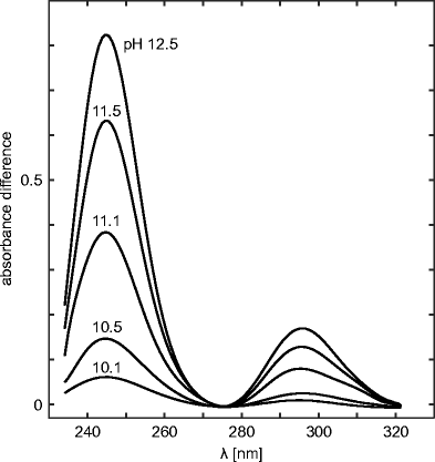
The departure spectra of actin equally a part of pH. Protein concentration is 0.17 mg/ml. The reference solution is of the same protein concentration at pH seven.3. Taken from Mihashi and Ooi (Mihashi and Ooi 1965)
Changes in absorption spectra, followed at ∼295 and/or ∼245 nm, indicate either ionization of exposed Tyr side-groups, when they occur at lower pH range, or ionization of buried residues at pH values sufficiently high to crusade denaturation of the protein and exposure of Tyr residues buried before the transition. Every bit a reference value of the pK a , which allows usa to consider it as loftier or normal, we can take a value found for the Tyr chromophore fully exposed to the solvent, e.yard., nine.76 for acetyl-Gly-Tyr-Gly-amide past NMR spectroscopy (Platzer et al. 2014).
Spectrophotometric titration can provide more than specific structural information. Mihashi and Ooi (1965) noted that abnormal Tyr residues remain even in half dozen M urea, even though such conditions unfolded helical segments of the protein. They concluded that the helical segments are located near the surface and Tyr located at these segments correspond i kind of buried residues. Simultaneously, deeply cached Tyr collaborate with other side-concatenation groups to maintain a rigid structure, presumably forming the core of the actin monomer. Urea destroys the helical construction of the molecule only is not strong enough to disrupt the interaction of deeply buried Tyr with other groups in the core. Add-on of guanidine-HCl unfolds not merely of the helical region but also of the region where the more stable aberrant Tyr residues are cached, perhaps consisting of hydrophobic amino acid residues. Classification of the abnormal Tyr into two kinds is consistent with this. Furthermore, the region containing the abnormal Tyr is distinguishable from a region that was disquisitional for polymerization, since the loss of polymerizability occurs before normalization of the Tyr by guanidine-HCl.
These titrations tin be combined with crystallography, as e.thou., for vipoxin neurotoxic phospholipase A2 (PLA2) (Georgieva et al. 1999). This basic poly peptide has a single polypeptide chain containing 122 residues, including 8 Tyr residues. Earlier crystallographic studies resulted in a three-dimensional construction, and immune the authors to determine solvent accessibility of the Tyr residues. Then, past spectrophotometric titration, they found three Tyr with a pKeff = x.45, iii with a pKeff = 12.17, and 2 with a pKeff = thirteen.23. Based on solvent accessibilities, Y117, Y75, and Y22 were identified as the first group, Y28, Y113, and Y52 every bit the 2d grouping, and Y25 and Y73 every bit the final group.
Derivative assimilation spectroscopy
Decision of Tyr exposure in proteins past second-derivative spectroscopy was proposed by Ragone et al. (1984), based on an earlier study of Servillo et al. (1982). This simple method examined Tyr and Trp in native proteins. Information technology is based on the observation that the second-derivative spectrum of N-Ac-Trp-NH2, between 280 and 300 nm (see Fig. 2) shows two maxima centered at 287 and 295 nm and two minima at 283 and 290.5 nm (Servillo et al. 1982; Ragone et al. 1984), and that they are only marginally affected past solvent polarity. In the same spectral region, North-Ac-Tyr-NHtwo exhibits a single minimum centered at 283.5 nm and ii maxima at 280 and 289.5 nm. Because intensities of Trp bands are much higher than those of Tyr bands, the second-derivative spectra of samples containing both more closely resemble that of Due north-Ac-Trp-NHtwo, even in the presence of loftier Tyr concentrations (Servillo et al. 1982).
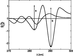
Second-derivative spectra of equimolar solutions of Due north-Ac-Trp-NH2 and North-Ac-Tyr-NHii dissolved in 6.0 Chiliad Gdn ⋅HC–0.05 Chiliad phosphate, pH 6.5. The spectrum of N-Ac-Trp-NH2 is identified by the two arrowsa and b, which indicate the peak-to-peak distances between the maximum at 287 nm and the minimum at 283 nm, and the maximum at 295 nm and the minimum at 290.5 nm, respectively. Taken from Ragone et al. (1984)
Ragone et al. (1984) focused on the ratio between two summit-to-peak distances, r p ≡a/b, with a and b defined in Fig. 2, in the second-derivative spectrum of Northward-Ac-Trp-NH2
$$ r_{p} \equiv \frac{a}{b} = \frac{A^{\prime\prime number}(\mathrm{287nm})-A^{\prime\prime number}(\mathrm{283nm})}{A^{\prime number\prime number}(\mathrm{295nm})-A^{\prime number\prime}(\mathrm{290.5nm})} $$
(1)
They found that r p is almost the aforementioned over a broad range of solvent polarities, with a mean value of 0.68 ±0.02. Considering the absorbance at any given wavelength of a mixture of the Trp and Tyr chromophores,
$$A = \epsilon_{\text{Trp}} \cdot C_{\text{Trp}} + \epsilon_{\text{Tyr}} \cdot C_{\text{Tyr}} $$
they derived the following equation for the second-derivative differences ΔA 1≡A 287−A 283 and ΔA 2≡A 295−A 290.5:
$$ \frac{\Delta A_{1}^{\prime\prime number}}{\Delta A_{two}^{\prime number\prime number}} = \frac{\frac{\Delta\epsilon^{\prime number\prime}_{ane,\,\text{Trp}}}{\Delta\epsilon^{\prime\prime}_{2,\,\text{Trp}}} + \frac{\Delta\epsilon^{\prime\prime}_{ane,\,\text{Tyr}}}{\Delta\epsilon^{\prime\prime number}_{2,\,\text{Trp}}} \cdot \frac{C_{\text{Tyr}}}{C_{\text{Trp}}}}{1 + \frac{\Delta\epsilon^{\prime\prime number}_{2,\,\text{Tyr}}}{\Delta\epsilon^{\prime\prime number}_{ii,\,\text{Trp}}} \cdot \frac{C_{\text{Tyr}}}{C_{\text{Trp}}}} \equiv \frac{{\mathcal{A}} x + {\mathcal{B}}}{{\mathcal{C}} ten + one} \equiv r_{d} $$
(ii)
where x≡C Tyr/C Trp, and definition of coefficients \({\mathcal {A}}\), \({\mathcal {B}}\), \({\mathcal {C}}\) clearly results from comparison of the two right terms in Eq. ii. Finally, in analogy to Eq. 1, defining r p , Eq. 2 may be considered as a definition of a similar quantity, given the symbol r d . Notation that if C Tyr = 0, so r d = r p , and the right-hand side of Eq. 2 can be written as
$$ r_{d} = \frac{{\mathcal{A}} ten + r_{p}}{{\mathcal{C}} x + 1} $$
(3)
which is a useful course if we have into account the experimentally verified independence of r p on solvent composition. However, Ragone et al. (1984) found that r d depends not only on the molar ratio x between Tyr and Trp, simply besides on the solvent composition. This dependence is a issue of the fact that the numerical values of coefficients \(\mathcal {A}\) and \(\mathcal {C}\) depend on the properties of the solvent. Investigating these dependencies, they showed that the numerical value of r d for a given Tyr/Trp ratio is determined by the molecular environs of tyrosine.
The second-derivative spectrum of proteins containing both Tyr and Trp residues shows the same general features observed for mixtures of Due north-Ac-Tyr-NH2 and N-Ac-Trp-NHii, i.eastward., two minima centered effectually 283 and 290.5 nm and ii maxima around 287 and 295 nm. Since the position of the peaks do not alter much after exposure to perturbing agents, Ragone et al. (1984) analyzed the second-derivative spectra of proteins in terms of the ratio betwixt the peak-to-summit distances a and b. They proposed the following equation to decide the caste of Tyr residue exposure to solvent in native proteins:
$$ \alpha = \frac{r_{due north} - r_{a}}{r_{u} - r_{a}} $$
(4)
where r n and r u are the numerical values of the ratio a/b adamant for the native and unfolded protein, respectively, and r a is the a/b value of a mixture, containing the same molar ratio of aromatic amino acids dissolved in a solvent with the same characteristics equally the interior of the protein, e.g. ethylene glycol. The fraction of Tyr residues exposed to solvent in several proteins with native structure, determined past second-derivative measurements, appears to be in good agreement with those available from crystallography or other methods (Ragone et al. 1984).
Second-derivative UV absorbance spectroscopy may be also used to decide pK a of Tyr side-chains in proteins (Breydo et al. 1997). The method monitors the decrease in intensity of the second-derivative of the spectrum at the isosbestic point corresponding to the transition betwixt cached and exposed non-ionized Tyr residues (284.2 nm). Existence of the isosbestic point (encounter an instance in Fig. iii) indicates that the transition from buried to exposed states of Tyr is a transition betwixt two discrete conformations of the protein. Hence, changes in the 2nd-derivative of the UV (SDUV) absorbance spectrum for a protein, at 284.2 nm, as a function of pH, reflects changes in the ionization state of the protein Tyr residues. An example of such titration curve is shown in Fig. iv.
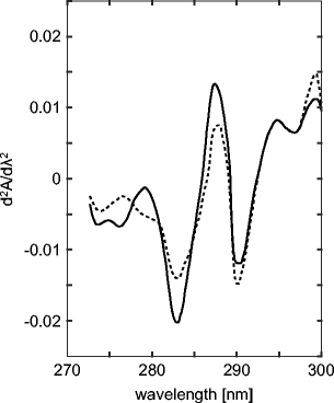
Isosbestic point at ∼284.2 nm in the second-derivative spectra of 2.3 μGrand native (50 mM Tris/acetate buffer pH 7.4; total line) and Gdn ⋅HCl denatured MM-creatine kinase (l mM Tris/acetate, 6 One thousand Gdn,HCl buffer pH 7.4; dotted line). Adapted from Leydier et al. (1997)
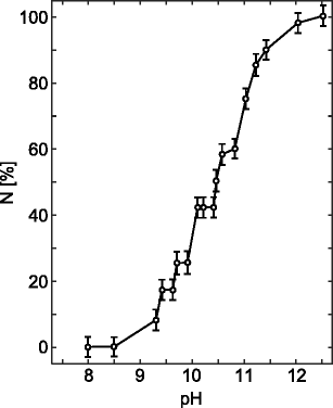
Titration curve of the Tyr residues in Carlsberg subtilisin obtained by the difference SDUV method, shown as % of ionized Tyr residues, N, vs. pH. Adapted from Breydo et al. (1997)
A modification of the above method was introduced for proteins with loftier levels of Trp residues (Tyr:Trp ratio less than ii, due east.g., for chymotrypsinogen and chymotrypsin). Breydo et al. (1997) analyzed such cases by using difference SDUV, i.east., by subtracting the spectrum of the same protein at the same concentration and at neutral pH from the spectrum recorded at a current pH.
The development of high-resolution ultraviolet absorbance spectroscopy permits the detection of the position of derivative peaks with a resolution approaching 0.01 nm. This encouraged (Lucas et al. 2006) to use the technique to investigate cation- π interactions in proteins. Their arroyo used high-chloride salt concentrations (Li +, Na +, and Cs +), chosen on the basis of their ionic radii to drive the diffusion of cations into interior regions of proteins where the cations collaborate with the aromatic amino acid side-bondage (Phe, Trp, Tyr). For each aromatic amino acid, they selected a tiptop in the SDUV spectrum to observe shifts every bit a function of table salt concentration. They expected to deduce local structure and dynamics of the eight selected proteins, albeit with varying degrees of exposure of detail side-chains. As command experiments, they investigated the ability of the three cations to shift the positions of aromatic absorbance peaks of the N-acetylated C-ethyl esterified amino acids, representing totally exposed side-chains. Here we focus on their results for Tyr residues.
Figure 5 shows the relative acme shifts for Northward-acetylated carboxyl ethyl ester of Tyr equally a role of cation type and concentration. Amino acids Due north-acetylated and esterified at the C-termini were chosen to minimize electrostatic interactions that could complicate interpretation of spectral changes due to cation- π furnishings between the salts and the aromatic ring of an amino acrid. The cations appear to induce quite dramatic height shifts in the Tyr spectra. Cs + causes a large red (positive) shift, Na + a small bluish (negative) shift, while Li + causes a large negative change, approximately equal in magnitude to the red shift effected by Cs +. These differences are also seen at concentrations of the guild of 0.25 Yard, suggesting that Tyr peak position is sensitive to both cation size or charge density and concentration.
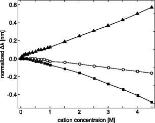
Peak shifts (Δλ) in the second-derivative UV absorption spectra for the model amino acid, N-acetyl-50-tyrosine ethyl ester, induced past the cations Li + (filled squares), Na + (open circles), and Cs + (filled triangles). The acme positions at each concentration point are normalized by subtracting the initial meridian position, which was 275.01 ±0.01 nm. Taken from Lucas et al. (2006)
Figure 6 illustrates the Tyr peak shift data for ribonuclease T1 (RNase T1, top), and human serum albumin (HSA, bottom), respectively. But these two of 8 proteins investigated past Lucas et al. (2006) are shown. The height shifts for RNase T1 are similar to those for the free amino acrid analog (Fig. 5). Increasing the Cs + concentration causes a cherry shift of the Tyr peak, while Na + causes an intermediate alter, and Li + a blue shift. Although the shifts are small, the similarity of these data to that of Northward-acetyl-l-tyrosine ethyl ester suggests that ane or more of the protein Tyr residues are located most the surface. The average solvent-attainable area for the Tyr residues of RNase T1 is relatively low, only the range of areas reveals that indeed some of the Tyr residues are near the poly peptide surface.
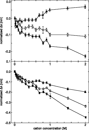
Tyr superlative shifts in the second-derivative UV absorption spectra of Ribonuclease T1 (elevation) and homo serum albumin (bottom), induced by the cations Li + (filled squares), Na + (open circles), and Cs + (filled triangles). The data are displayed equally in Fig. 5, with initial peak positions of 277.61 ±0.01 nm for RNAse T1, and 278.82 ± 0.01 nm for HSA. Error bars represent the standard deviation of the mean for three experiments for each salt. Taken from Lucas et al. (2006)
The top shifts for HSA (top of Fig. vi) are similar to those for the complimentary amino acid analog (Fig. 5) in the case of Li + and Na + cations, whereas for Cs + trends in the 2d-derivative UV spectra are contrary to the command information for free Tyr. This suggests that the phenyl rings are buried, or that the protein construction has been perturbed. The three cations induce a large bluish shift in the HSA Tyr peak (bottom of Fig. half dozen). The authors concluded these changes betoken that hydrogen bonding dominates the observed pinnacle shifts in the presence of all three cations. The Tyr residues of this protein have a relatively high degree of solvent exposure compared to Tyr residues in other proteins studied by them.
Kinetic aspects of Tyr exposure and/or pK a determination, revealed by stopped-flow spectrometry
Kuwajima et al. (1979) developed a stopped-menstruation technique for zero-time spectrophotometric titration of Tyr residues in the native or in the completely alkaline-denatured state applicable to proteins that undergo relatively fast element of group i conformational transition in the pH region of Tyr ionization.
When pH of a protein solution is suddenly raised sufficiently high in a stopped-flow experiment, two kinds of changes occur: (i) Accessible Tyr are ionized (this procedure is completed inside dead time of stopped-menses apparatus, i.e., ∼1 ms); and (2) A reversible time-dependent change in conformation (which can make other Tyr accessible, and then ionized) is induced, which proceeds to an equilibrium endpoint that, in plow, depends on pH. If these changes are observed using UV assimilation, the full change can be represented every bit a sum of three contributions (Kuwajima et al. 1979)
$$ {\Delta} A = {\Delta} A_{\text{ion}} + {\Delta} A_{\text{conf}} + {\Delta} A_{\mathrm{conf+ion}} $$
(5)
One tin only detect contributions of the 2d and tertiary term in the correct-paw side of Eq. 5 to the time-dependent absorbance changes, ΔA conf and ΔA conf+ion, provided that the conformational change caused past the pH-spring is not too rapid for stopped-flow experiments. When this condition is satisfied, then extrapolation of the observed absorption changes to cypher time gives information about the ionization equilibrium in the native land at the alkali metal pH. Determining this absorbance modify relative to aught time as a function of the final alkaline pH leads to a titration curve, which was denoted by Kuwajima et al. (1979) every bit the zero-time titration curve for the native state. Similar assay for pH-jumps from highly element of group i pH to lower values resulted in the zero-time titration curve for the purely element of group i-denatured protein.
Kuwajima et al. (1979) applied this to bovine α-lactalbumin, characterized past occurrence of fast alkaline denaturation, which makes determination of the ionization behavior of tyrosines by usual spectrophotometric techniques practically impossible.
Offset, it was necessary to separate contributions to the spectra due to ionization of the Tyr residues from contributions due to alkaline denaturation itself. To obtain the spectrum caused by alkaline denaturation, Kuwajima et al. carried out pH-jump experiments from pH 11.six to pH 8.0. At pH eight.0 the protein is in the native (N) state and all its Tyr residues are protonated. At the initial pH of xi.6, the protein is in the element of group i-denatured (D) state. With a pH-jump from xi.6 to 8.0 in a stopped-flow spectrometer, protonation of the Tyr residues exposed to the solvent at the starting pH, is instantaneously lost in the dead time of the experiment leaving simply the absorbance change respective to ΔA conf in the kinetic traces. This immune Kuwajima et al. to determine kinetic amplitudes observed by the pH-jump from 11.6 to viii.0, equally a function of the incident wavelength. They showed that at 298 nm the departure absorption in the spectrum is essentially zero. Thus, the absorption change at 298 nm can be taken as a measure of Tyr ionization.
Figure 7 shows the typical kinetic traces for absorption at 298 nm from stopped-menstruum experiments, one for a pH-bound from pH 5.five to 11.three, and the other from pH xi.viii to 10.two. These were analyzed using a first-order rate law:
$$ A(t)-A_{o} = \left( A_{\infty} - A_{o}\right) \cdot \left( 1-\exp(-kt)\correct) $$
(6)
where A(t) is the absorption at time t after the pH-spring, \(A_{\infty }\) and A o are the values at space and at naught time, respectively, and k is the get-go-order charge per unit constant. The divergence \(A_{\infty }-A_{o}\) represents the desired "kinetic amplitude" of the observed processes (\({\Delta }\epsilon _{\mathrm {298}}^{f}\) for forward, and \({\Delta }\epsilon _{\mathrm {298}}^{r}\) for reversed pH-jump, respectively).
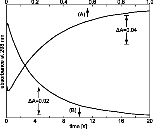
Typical time-courses of the absorption changes at 298 nm after a pH-leap at 0.5 K Gdm-HCl and 25.0 ∘C : a forward pH-spring from pH 5.5 to xi.3; and b reversed pH-jump from 11.8 to 10.2. Protein concentration is ca. 0.05 %. Taken from Kuwajima et al. (1979)
The pH-dependence of the equilibrium divergence extinction coefficient at 298 nm, \({\Delta }\epsilon _{\mathrm {298}}^{\text {eq}}\), gives the equilibrium ionization curve of the alkaline denaturation. The transition from low pH (where the construction is native) to high pH (fully denatured state) exhibits a transition zone when both native and alkaline denatured species participate to the ionization equilibrium. Assuming a two-land transition, \({\Delta }\epsilon _{\mathrm {298}}^{\text {eq}}\) can exist related to the fractions of the native, (f N ), and denatured, (f D ), species, and the degrees of Tyr ionization in both species, (α Due north, i and α D, i) equally expressed past the following:
$$ {\Delta}\epsilon_{\mathrm{298}}^{\text{eq}} = {\Delta}\epsilon_{\mathrm{298}}^{\mathrm{0}} \sum\limits_{i=1}^{n} \left( f_{N} \alpha_{\mathrm{N,\,i}} + f_{D} \alpha_{\mathrm{D,\,i}}\correct) $$
(vii)
where \({\Delta }\epsilon _{\mathrm {298}}^{\mathrm {0}}\) is the extinction coefficient for ionization of a unmarried Tyr residue, and due north is the number of Tyr residues in the protein (iv in the case of α-lactalbumin).
When α N, i differs significantly from α D, i at the final pH in pH-jump experiments, the conformational change caused by the pH-spring leads to a change in Tyr ionization. This can be observed as a time-dependent absorption change of ΔA conf+ion at 298 nm, following a pH-jump from an initial value (∼5.v or ∼11.8) to some final pH, obeying the alkaline-transition region. Resulting amplitudes, \({\Delta }\epsilon _{\mathrm {298}}^{f}\) and \({\Delta }\epsilon _{\mathrm {298}}^{r}\), plotted against the final pH are shown in Fig. 8. The bell-shaped features of the plots are expressed past:
$$ {\Delta}\epsilon_{\mathrm{298}}^{f} = {\Delta}\epsilon_{\mathrm{298}}^{0} f_{D} \sum\limits_{i=1}^{four} \left( \alpha_{\mathrm{D,\,i}} - \alpha_{\mathrm{N,\,i}}\right) $$
(8)
and
$$ {\Delta}\epsilon_{298}^{r} = {\Delta}\epsilon_{298}^{0} f_{Northward} \sum\limits_{i=1}^{4} \left( \alpha_{\mathrm{N,\,i}} - \alpha_{\mathrm{D,\,i}}\right) $$
(nine)
where f N , f D , α Due north, i, and α D, i refer to the terminal pH. From Eqs. seven–ix, and the status f N + f D = 1, one obtains
$$ {\Delta}\epsilon_{298}^{\text{eq}} - {\Delta}\epsilon_{298}^{f} = {\Delta}\epsilon_{298}^{0} \sum\limits_{i=i}^{4} \alpha_{\mathrm{N,\,i}} $$
(10)
and
$$ {\Delta}\epsilon_{298}^{\text{eq}} - {\Delta}\epsilon_{298}^{r} = {\Delta}\epsilon_{298}^{0} \sum\limits_{i=1}^{iv} \alpha_{\mathrm{D,\,i}} $$
(11)
These two quantities correspond to the zero-time difference extinction coefficients of Tyr ionization after the pH-jump. They practise not include any contributions from the conformational transition. Thus, they can be used to plot the zero-time titration curves of tyrosines in the purely N and the purely D states, respectively.
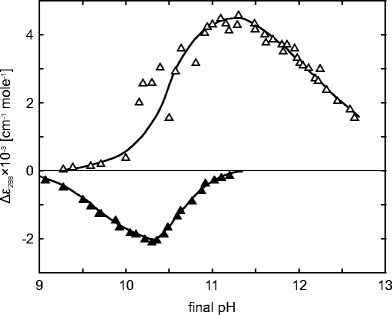
Dependence of \({\Delta }\epsilon _{\mathrm {298}}^{f}\) (empty triangles) and \({\Delta }\epsilon _{\mathrm {298}}^{r}\) (full triangles) on final pH. Taken from Kuwajima et al. (1979)
Bold contained ionization of the tyrosines, justified by the relatively loftier ionic strength of the solutions, the degrees of Tyr ionization tin can be related to equilibrium ionization constants K Northward, i and One thousand D, i, respectively and hydrogen ion activity \(a_{H^{+}}\):
$$ \alpha_{\mathrm{N,\,i}} = \frac{K_{\mathrm{N,\,i}}}{K_{\mathrm{N,\,i}}+a_{H^{+}}} \quad \text{and} \qquad \alpha_{\mathrm{D,\,i}} = \frac{K_{\mathrm{D,\,i}}}{K_{\mathrm{D,\,i}}+a_{H^{+}}} $$
(12)
These last expressions can be used in Eqs. 10 and 11 to fit theoretical predictions to experimental observations, resulting in values of equilibrium ionization constants defined by Eq. 12. The values of \({\Delta }\epsilon _{\mathrm {298}}^{0}\) were estimated from \({\Delta }\epsilon _{\mathrm {298}}^{0}\) at pH 12, where all the Tyr are fully ionized, since \({\Delta }\epsilon _{\mathrm {298}}^{0}\) of the protein completely denatured past half-dozen Grand Gdn ⋅HCl is almost the same every bit that for 0.5 M Gdn ⋅HCl at pH 12.
The effect is that, in the denatured state, all Tyr have pK a equal to 10.3, whereas in the native land they are: pK a, Tyr18 = 11.8, pK a, Tyr36 = xi.8, pK a, Tyr50 = 12.vii, and pK a, Tyr103 = ten.5 (Kuwajima et al. 1979).
Protein structure changes measured by tyrosine fluorescence
Tyr exhibits substantial fluorescence, and the high environmental sensitivity of its emission, making it a useful natural probe for studying construction and dynamics of proteins. Intrinsic fluorescence of proteins also results from excitation of two other effluvious amino acids, Trp and Phe. Because of the ascendant emission of Trp, fluorescence of Tyr can normally be routinely observed in proteins that lack Trp residues (Lakowicz et al. 1995). Notwithstanding, as noted past Edelhoch et al. (1969), in some proteins, fluorescence of both Trp and Tyr can be observed simultaneously. 1 such protein is the parathyroid hormone, PTH, an of import endocrine regulator of calcium and phosphorus concentration in extracellular fluid.
Bovine PTH (BPTH) is a single polypeptide chain of 83 amino acids and only i Trp and 1 Tyr (Edelhoch and Lippoldt 1969). The emission spectrum of BPTH at pH vi.i (excitation at 270 nm) is depicted in Fig. nine. The fluorescence of Tyr is visible as a pronounced shoulder at 300 nm. The pinnacle of Trp fluorescence in this polypeptide occurs at 347 nm. Position of the elevation and the relative quantum yields of the two chromophores, make it possible to detect a distinctive band due to Tyr emission (Edelhoch et al. 1969). When BPTH is excited at 290, instead of 270 nm, 65 % of the Tyr, but only ten % of the Trp emission is lost (Edelhoch et al. 1969).
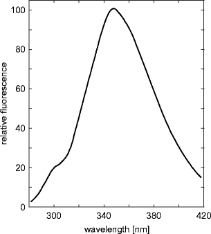
Fluorescence spectrum of PTH, pH 6.1, 0.08M KCl, 0.02M lysine, with excitation at 270 nm. Adjusted from Edelhoch et al. (1969)
The emission spectrum of BPTH in the range of 280 to 400 nm is almost superimposable on the emission spectrum of Trp-Glyiv-Tyr (Edelhoch and Lippoldt 1969), suggesting that the boilerplate distance distribution betwixt the two chromophores is similar in both peptides (Edelhoch et al. 1969). A more precise estimate of the distance between the 2 chromophores can be determined from the extent of Trp fluorescence quenching resulting from Tyr ionization.
The quenching of Trp fluorescence emission past radiationless free energy transfer (FRET) to ionized Tyr has been shown to exist mutual in proteins in the alkaline pH range (Edelhoch and Lippoldt 1969; Steiner and Edelhoch 1963). Effigy ten shows the effect of pH on fluorescence of Tyr and Trp in BPTH. Both emissions are normalized to the aforementioned value at neutral pH. It can be noted that Trp fluorescence is quenched by 30 % if the bend is extrapolated to 100 % ionization of the Tyr. A proportionality between Trp quenching and Tyr quenching, property in the full pH range of Tyr ionization, suggests that the quenching of Trp fluorescence is due solely to energy transfer to the ionized Tyr residual (Edelhoch and Lippoldt 1969). A much smaller degree of Trp quenching (10 %) of PTH by ionized Tyr is observed in 4.half dozen G guanidine solution (see Fig. 10). These results can exist interpreted based on experiments with Trp-Gly\(_{0\leftrightarrow four}\)-Tyr, which showed that the degree of quenching of Trp emission by Tyr ionization decreased regularly with increasing chain length, and vicious to nigh 40 % in Trp-Gly4-Tyr (Edelhoch and Lippoldt 1969). Judging from the decrease in quenching efficiency in the serial Trp-Gly\(_{0\leftrightarrow 4}\)-Tyr, the inter-residue distance in BPTH in guanidine-HCl is presumably greater than in water (Edelhoch et al. 1969; Edelhoch and Lippoldt 1969).
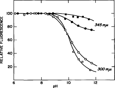
The alkaline dependence of tyrosyl (300 nm) and tryptophan (345 nm) fluorescence of PTH in aqueous solution (circles) 0.090M KCl, 0.02M lysine and in four.vi M guanidine-HCl (squares). Excitation at 270 nm at 25 ∘C. Taken from Edelhoch et al. (1969)
Insulin is a tryptophan-complimentary peptide hormone produced by β-cells in the pancreas. Information technology is essential for regulating homeostasis of blood glucose levels (Hua 2010), and used to treat insulin-dependent diabetics. It is a globular protein composed of two peptide chains, A and B, with 21 and 30 amino acid residues, respectively. The molecule is linked by iii disulphide bridges (two inter-chain: A7-B7 and A20-B19, and 1 intra-A-concatenation: A6-A11). Four of the residues are Tyr: A14, A19, B16, and B26 and three are Phe: B1, B24, and B25.
Bekard and Dunstan (2009) monitored intrinsic Tyr fluorescence of bovine insulin in 0.1 % (v/v) HCl, to investigate its partial unfolding and fibrillation. The pH of solution (1.nine), being well below the pKa of both footing state (pK a = ∼ten) and excited state (pK a = ∼4.2) of the chromophore, ensured that deprotonation of the hydroxyl grouping and subsequent formation of tyrosinate or Tyr-carboxylate hydrogen bonds could be excluded. Fluorescence of Tyr residues in insulin was monitored using excitation of 276 nm, with emission measured at 303/305 nm for kinetic studies, or scanned in the interval 280-500 nm for equilibrium studies.
Figure xi (left) presents the Tyr fluorescence spectra of insulin incubated nether atmospheric condition favoring fibrillation equally a role of fourth dimension and shows a subtract in emission intensity with time. Simultaneously, λ max of the ring is constant at 305 nm. Plotting the emission intensity at 305 nm against fourth dimension (shown in the inset) gives a sigmoid bend. Taken together, data presented in Fig. 11 (left) clearly shows a fast decrease in emission intensity later ∼2 h incubation, and reaching quasi-equilibrium state at 12 h. The authors discussed several possible catalysts for the observed Tyr fluorescence quenching preceding insulin fibrillation (Bekard and Dunstan 2009).
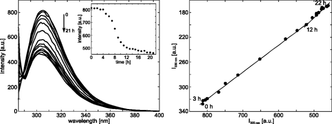
Left: Fluorescence emission spectra of Tyr during insulin aggregation initiated by incubation under conditions favoring fibrillation. Spectra registered at 1-h intervals are shown. The inset shows a sigmoidal change observed in the emission intensity at 305 nm equally a part of fourth dimension; Correct: Phase diagram obtained from Tyr fluorescence data that reveal the existence of structural intermediates on thermal denaturation of insulin. Taken from Bekard and Dunstan (2009)
Farther insight into the machinery of insulin fibrillation is provided by a phase diagram of Tyr fluorescence intensity at 330 and 303 nm (Fig. eleven right). The stage diagram method consists of building up a correlation between two fluorescence intensities (extensive parameters in general) at two wavelengths, I(λone) and I(λ2), under dissimilar experimental conditions, thereby forcing the protein to undergo structural transformations (Ahmad et al. 2003). When applied to protein folding, the relation I(λ1) = f[I(λ2)] is linear if changes in the protein environs lead to a two-land transition between two different conformations. The presence of a number of linear segments indicates the sequential structural transformations, where each linear portion of the I(λ1) = f[I(λ2)] plot describes an individual all-or-none transition (Ahmad et al. 2003).
Bekard and Dunstan (2009) applied phase diagram analysis to a study of the effect of temperature on insulin betwixt 10 and 95 ∘C. The resulting fluorescence phase plot (Fig. 11 right), exhibits three linear segments suggesting two intermediate states in the transition from native to provisionally denatured insulin.
VanderMeulen and Govindjee (1977) suggested the molecular mechanism of photophosphorylation in chloroplasts. They measured Tyr fluorescence polarization of ATP synthase, excited at 280 nm in the presence of cations, ADP and P i . ATP synthase is an enzyme that provides energy by synthesizing adenosine triphosphate (ATP).
Figure 12 shows the polarization data for Tyr fluorescence in ATP synthase to monitor possible functional changes in its conformation induced by substrates or cofactors of phosphorylation. The improver of divalent cations results in a 20 % increase in polarization of Tyr fluorescence. MgCl2 is somewhat more efficient than CaCl2 in producing this effect at low (2.5-20 mM) concentrations just this disappears at xxx mM. With KCl, the resulting changes are much smaller in the same concentration range. The addition of 2.5-20 mM MgClii had little or no upshot on either the UV absorption (280 nm) or fluorescence spectra, suggesting that the observed salt-induced changes in polarization of ATP synthase fluorescence are not due merely to a decrease in the lifetime of Tyr fluorescence. The authors suggest these changes are due to an altered protein construction in which, for example, the mobility of one or more Tyr residues is reduced.
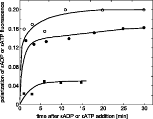
The consequence of various salts on polarization of Tyr fluorescence in ATP synthase. The medium contained 25 mM tricine-NaOH (pH 8.2) and 3.6 μM protein. Taken from VanderMeulen and Govindjee (1977)
Our next example is a study of fluorescence polarization decay of Tyr in lima bean trypsin inhibitor (LBTI), an 83 amino acid protein with vii disulphide bonds (Nordlund et al. 1986). Trypsin inhibitor is a serine protease inhibitor that reduces the biological activity of trypsin, an enzyme involved in the hydrolysis of proteins during digestion.
Nordlund et al. (1986) investigated the fluorescence anisotropy decay of the single Tyr-69 in LBTI to gain insight into the time-scale of structural fluctuations and motions of this protein. Tyr fluorescence lifetime, obtained from a unmarried-exponential, is 620 ±50 ps. The results of anisotropy decay are interpreted by comparison with the anisotropy decay of N-Ac-Tyr-NH2 in a viscous medium. In each case, experimental results were fitted to
$$ r(t) = r_{0} \mathrm{Eastward}^{-t/\phi} \left[a\mathrm{e}^{-t/\phi_{i}} + (1-a)\right], $$
(xiii)
where ϕ is the rotational correlation time for the whole protein, ϕ i is that for the internal motion, and a is the fraction of the total anisotropy that decays as a effect of the internal motion. Assuming time zero as the fourth dimension corresponding to the heart of the excitation pulse, Norden et al. fit the fluorescence anisotropy disuse of LBTI to a dual-exponential form, with time constants of virtually 40 ps and 3 ns. The nanosecond component is consistent with rotation of the unabridged poly peptide molecule. The 40-ps component demonstrates that the Tyr has much greater liberty of motion.
Structural analysis of proteins measured by linear dichroism of tyrosine residues
LD provides data about the average orientation of the Tyr residues in proteins. Here we review a study of the Rad51-Dna filament (Reymer et al. 2009), a homologue of Escherichia coli RecA. Rad51 is a protein involved in repair of double-strand breaks in DNA in eukaryotic cells (Baumann and West 1998). Rad51 and RecA form extended filaments on DNA and promote the Deoxyribonucleic acid strand exchange in recombinant DNA repair (Cox 2009). Combining molecular modeling with LD of the human Rad51-dsDNA-ATP complex in solution (HsRad51-dsDNA-ATP), Nordén et al. (Reymer et al. 2009) proposed a model corresponding to the final product of the recombination reaction. Their experimental approach, called site-specific linear dichroism (SSLD), is based on molecular replacement of private aromatic residues in the protein with other aromatic residues, providing angular orientations of the replaced residues relative to the filament axis.
The HsRad51 lacks Trp, but contains ten Tyr residues that were replaced, ane at a fourth dimension, past Phe, using site-directed mutagenesis. The Tyr to Phe modification corresponds to a structural elimination of an oxygen atom, and is expected to leave the poly peptide structure unchanged. This was confirmed by comparing CD spectra of the wild-blazon (WT) and mutated proteins. The difference LD spectrum (SSLD) betwixt the WT and modified proteins, was considered to represent the LD spectrum of the replaced residue itself, after appropriate normalization for extinction coefficients of Tyr and Phe, and correction for the groundwork.
Figure thirteen left presents experimental flow LD spectra of the WT HsRad51-DNA-ATP filaments and two modified nucleoprotein filaments, in which Tyr was replaced by Phe at residues 205 and 228. Figure 13 correct shows two SSLD spectra for corresponding Tyr residues and the baseline correction spectrum.
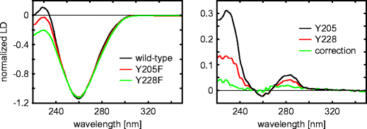
Left: Experimental flow LD spectra of wild-type and two selected modified HsRad51 nucleo-protein complexes; Right: resulting SSLD spectra for the respective Tyr residues. Adapted from Reymer et al. (2009)
There are singled-out variations in the spectra of modified poly peptide complexes, compared to WT protein solitary. The nearly obvious differences are centered at 230 nm, only the spectra also differ around 280 nm. These wavelengths correspond to the L a and Fifty b transition moments, respectively, of Tyr.
Assuming that the structures of nucleo-protein filaments formed by WT and modified proteins are comparable, the SSLD spectrum of the particular substituted residue tin can be computed as the differential spectrum of the LD spectrum of the WT and mutated protein complexes. The SSLD spectrum of the substituted remainder may be used to make up one's mind orientation coordinates of the replaced remainder chromophore with respect to the filament axis oriented along menses lines of the solvent in the flow LD experiment.
The angular orientation β of the transition moments, for each substituted Tyr residue relative to the helix axis of the nucleo-protein filament, tin be determined from the reduced LD if the orientation gene South in Eq. 6 of Part one is known. S is the degree of orientation of the filamentous complex achieved in the catamenia LD experiment. The value of this parameter was computed by Nordén et al. (Reymer et al. 2009), assuming that the orientation of DNA in the complex with Rad51 is like to that in the RecA-DNA complexes assessed from small-bending neutron handful (Nordén et al. 1992). Using this arroyo, the orientation angles for eight of the ten Tyr residues of HsRad51 were determined from the SSLD spectra. Two athwart orientations, the L a and Fifty b transition moments, were reported for each Tyr. The meaning of these orientations is explained past referring to Fig. 14. For Tyr205 and Tyr228 (the SSLD spectra are shown in Fig. thirteen) the Fifty a and Fifty b angles are 43 ∘ and 41 ∘, and 28 ∘ and 30 ∘, respectively.
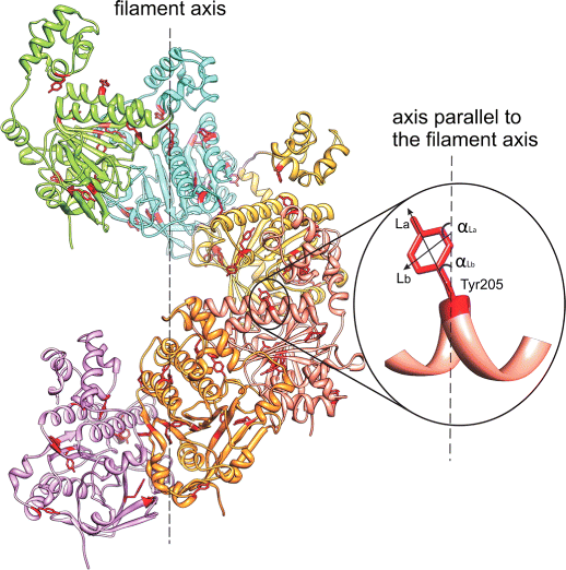
Molecular model of the HsRad51 helical filament showing the angular orientations of transition moments L a and 50 b of Tyr-205 balance, relative to the filament axis. Taken from Reymer et al. (Reymer et al. 2009)
The published results (Reymer et al. 2009) were discussed in a later publication (Fornander et al. 2014) in the context of the evidence that the spectral shifts of the Tyr chromophore, p-cresol, are due to ecology changes, mainly a result of its power to form hydrogen bonds. The observed clear red- (Tyr54 and Tyr216 of HsRad51) and blue shifts (Tyr205 and Tyr228) of the absorption bands of L a transition were interpreted (Fornander et al. 2014) by referring to the hydrogen bonding capabilities suggested by considering previously developed fragments of the Rad51 protein construction based on crystallography and NMR (Aihara et al. 1999; Pellegrini et al. 2002; Conway et al. 2004).
Tyrosines in near-UV CD spectrometry of proteins
Tyrosyl CD bands of proteins in the near-ultraviolet may be used to study their tertiary structure in solution. However, this is complicated by Trp, Phe, and Cys residues absorption bands in the same wavelength range. Horwitz et al. (1970). Even in proteins lacking Trp, the Tyr CD bands are not easily identified, nor can their intensities exist accurately assessed, because of ambivalence due to disulphide contributions. One instructive example is given by ribonuclease A (RNase-A) where the 275-nm CD band tin exist mostly attributed to Tyr. Alien interpretations depend on whether cached or exposed Tyr side chains can generate the observed CD ring (Horwitz et al. 1970; Simpson and Vallee 1966; Simmons and Glazer 1967).
Bovine pancreatic RNase-A contains 6 Tyr residues, three Phe residues, no Trp, and iv conserved disulphide bonds (Howlin et al. 1989; Chatani et al. 2002) (see Fig. 15). 3 RNase-A tyrosines, Tyr25, Tyr92, and Tyr97, are buried and three, Tyr73, Tyr76, and Tyr115, are exposed to solvent.
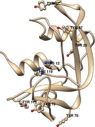
A ribbon diagram of bovine pancreas RNase-A showing the six tyrosines. His12 and His119 are also shown to betoken the location of the active site. Generated by USCF Chimera [REF: Pettersen EF, Goddard TD, Huang CC, Couch GS, Greenblatt DM, Meng EC, Ferrin TE (2004) J Comput Chem 25:1605] from the structure of RNase A by Howlin et al. (1989).
Using assimilation and CD spectra of tyrosine and three of its derivative, registered at 298 and 77 ∘Chiliad, and referring to the vibronic transitions of p-cresol, Horwitz et al. (1970) analyzed assimilation and CD spectra of RNase-A, besides at 298 and 77 ∘One thousand. They intended to decide the relative contributions of the diverse types of Tyr residues. They identified 0-0 transition for two tyrosines at 286 nm, for one tyrosine at 289 nm, and for three tyrosines at 283 nm, in the absorption spectrum at 77 ∘Thousand. Spectral positions of the latter indicated they are solvent-exposed. In the CD spectrum of RNase-A, obtained at 77 ∘K, they identified well-resolved bands at 282 and 276 nm, and shoulders at 288.five, 267.5, 261, and 255 nm. The first three were assigned to Tyr, and the last three to Phe. Horwitz et al. (1970) found no CD band at 286 nm, corresponding to ii Tyr residues with 0-0 absorption at this wavelength. They concluded that the contribution from these Tyr residues is insignificant.
Recently, much more extensive data on Tyr side-concatenation contributions to the spectra (both in the near- and far-UV range) of RNase-A was presented by Woody and Woody (2003). They measured and computed CD spectra of wild-blazon (WT) and six Tyr to Phe mutants. Comparing of WT and mutated RNase-A CD spectra, both experimental and theoretical, get in possible to resolve roles of individual Tyr to the CD spectra. Here we limit the discussion to experimental CD spectra in the near-UV range.
Comparing the far-UV CD spectra of the Tyr →Phe mutants with the spectrum of the WT protein, at ii ∘C and pH 7, Woody and Woody (2003) ended that the secondary structures of the mutants are not significantly perturbed. Thermal unfolding experiments of WT RNase-A and the mutants revealed that melting temperatures of the mutants vary only slightly from that of the WT. The largest deviations, [T k (mutant) −T m (WT)], were observed for two of the mutations at buried Tyr sites: −5.0 ∘C for Y25F, and −9.7 ∘C for Y97F.
3 exposed Tyr in RNase-A titrate with a pK a of ∼nine.9 and 3 buried tyrosines have elevated pK a values (Shugar 1952; Tanford et al. 1956), thus Woody and Woody expected to find a difference in the CD spectra due to contributions of the three exposed and three buried Tyr past raising the pH. The pH difference spectra of the mutants and the WT RNAse are shown in Fig. sixteen left. The deviation spectrum for the WT has a negative band at 295 nm and an enhanced positive band at 245 nm. The mutants of the three cached Tyr (Y25F, Y92F, and Y97F) and one exposed Tyr (Y76F) all exhibit pH departure spectra similar to the WT. The mutants of two exposed Tyr (Y73F and Y115F) exhibit macerated 295-nm negative bands and, instead of positive bands, negative bands are observed at 245 nm. Assuming that disulphide and peptide bond contributions to the CD spectra do not change essentially with changing pH, the dual differences in the ellipticities ΔΔ𝜖 295≡Δ𝜖 295(pH xi)−Δ𝜖 295(pH seven) and similarly defined ΔΔ𝜖 245 probably represent contributions from Tyr lone.
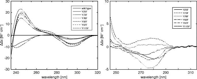
Left: The pH difference CD spectra (pH xi.3 and 7.0, two ∘C) of RNase A wild-type and Tyr →Phe mutants.; Correct: Experimental near-UV CD difference spectra (2 ∘C) of RNase-A wild-type minus the mutants at pH 7.0. Taken from Woody and Woody (2003)
Analyzing the values of ΔΔ𝜖 295 and ΔΔ𝜖 245, Woody and Woody noted that mutation of two of the buried Tyr (Y25F and Y97F) and one of the exposed Tyr (Y76F) slightly perturb the ΔΔ𝜖 295 and ΔΔ𝜖 245 values from those of the WT. This suggests that the contributions of these Tyr to the 277- and 240-nm bands are minor, merely non negligible. Two subsequent mutants (Y73F and Y115F) show a substantial reduction in the ΔΔ𝜖 295 values and exhibit negative ΔΔ𝜖 245 values, understood as indicating that these two exposed Tyr are a significant source of the 277- and 240-nm bands.
The right panel in Fig. sixteen shows the Tyr contributions to 277-nm CD difference spectra for WT and the mutants. The mutants at two of the exposed Tyr sites (Y73F and Y115F) take reduced negative intensity in the near-UV CD band. This results in more negative ΔΔ𝜖 277≡Δ𝜖 277(WT)−Δ𝜖 277(mutant) values than those from Y25F and Y92F. The mutants Y76F and Y97F show positive ΔΔ𝜖 277 values. These spectra indicate not only that the two exposed Tyr (73 and 115) contribute significantly to the CD at 277 nm, but as well that the contribution of buried Tyr are not negligible. Some other interesting result is the ΔΔ𝜖 286 (defined in analogy to ΔΔ𝜖 277) value for the WT-Y92F is negative and the value for the WT-Y97F is positive. Thus, the almost-UV CD contributions of Tyr92 and Tyr97, two cached tyrosines, are comparable in magnitude, merely opposite in sign. Co-ordinate to Woody and Woody (2003), this explains why Horwitz et al. (1970) observed no CD band at 286. It was a issue of the canceling contributions of Tyr92 and Tyr97.
Resonance Raman scattering from tyrosine residues
Nosotros now nowadays several applications where the determination of the position and relative intensities of a closely spaced pair of Raman lines at ∼850 and ∼830 cm −1 due to Tyr is used. This is an example of a more than general phenomenon known every bit the Fermi resonance doublet (Larkin 2011).
Using continuous-moving ridge excitation at 244 nm, Couling et al. (1997) utilized the intensity ratio, R 244 = I 834/I 855 to investigate the environment of Tyr residues in native and denatured barnase. Barnase is a bacterial ribonuclease, consisting of 110 amino acids. It has three Trp (35, 71, 94) and 7 Tyr (xiii, 17, 24, 78, 90, 97, 103) residues (Fersht 1993). In the denatured form, all 7 Tyr residues are exposed to the solvent. In the folded state, ii, Tyr13 and Tyr17, remain exposed to solvent, while the other five are buried in hydrophobic cores (Matouschek et al. 1992).
Figure 17 shows Fermi-resonance doublets for denatured and folded wild-type barnase. In principle, one expects resonance Raman signals from both the Trp and Tyr residues. Furthermore, these features will exist the average for the 3 Trp and seven Tyr residues. The Fermi-resonance doublet seen at 834/855 cm −i is the average of all seven Tyr, so care is needed if the change in intensity ratio, R 244 = I 830/I 850, of the doublet is to yield quantitative information. In the denatured state, all seven Tyr residues are solvent-exposed, so the R 244, from intensities in the Figure, is R 244(denatured)=0.67±0.09, whereas for folded barnase,
$$R_{\mathrm{244}}(\text{folded}) = \mathrm{one.sixty\pm 0.16} = \frac{2}{vii}\cdot \mathrm{0.67} + \frac{5}{seven}\cdot R_{\mathrm{244}}(\text{buried}) $$
which yields 1.97 as the ratio for buried Tyr residues. A general formula for R 244 to exist observed in barnase with mole fraction x e of exposed Tyr and mole fraction x b of cached Tyr is thus
$$R_{\mathrm{244}} = i.97\cdot x_{b} + 0.67 \cdot x_{eastward} \qquad \text{with} \qquad x_{e}+x_{b}=1$$
Subsequently, one tin use the Couling et al. (1997) linear relation betwixt R 244 and enthalpy of hydrogen bail formation ΔH, obtained from values of the Tyr Fermi-doublet intensity ratio, R 244 = I 830/I 850, for the model chemical compound p-cresol in solvents of varying H-bond acceptor strengths,
$$R_{\mathrm{244}} = 0.31 \cdot \left( -{\Delta} H\right) + 0.19$$
where ΔH is in units of kcal ⋅mol −1. Accordingly, one may estimate average enthalpies of hydrogen bonding for exposed and buried Tyr residues in barnase. From R 244(exposed)=0.67, one gets ΔH = −ane.55 kcal ⋅mol −one, whereas from R 244(buried)=i.97, ane gets ΔH = −v.74 kcal ⋅mol −ane.
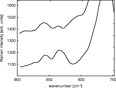
Resonance Raman spectra of barnase in the range 750-890 cm −1 obtained with excitation wavelength 244 nm, for denatured wild-blazon barnase (pH 1.5, top) and folded wild-type barnase (pH half dozen.3, bottom). The spectra have been displaced for clarity. Adapted from Couling et al. (1997)
A 2d example is decision of the pK a of Tyr in an eleven-remainder peptide fragment from transthyretin by Pieridou and Hayes (2009). Transthyretin (TTR) is a highly conserved homotetrameric protein, synthesized mainly past the liver and the choroid plexus of brain (Vieira and Saraiv 2014). It is a serum and cerebrospinal fluid carrier of the thyroid hormone thyroxine and retinol-binding protein bound to retinol. Originally, TTR was called prealbumin considering it runs faster than albumin on electrophoresis gels.
The investigated xi-residue fragment has the sequence Tyr-Thr-Ile-Ala-Ala-Leu-Leu-Ser-Pro-Tyr-Ser, which is located at positions 105 to 115 along the main chain of TTR, TTR(105-115). This peptide fragment was shown to grade amyloid fibrils, and hence may serve as a model system for studies of fibril formation in general (Gustavsson et al. 1991).
Pieridou and Hayes (2009) measured resonance Raman spectra of the TTR(105-115) peptide in solution as a function of pH, using excitation at 239.five nm, which is resonant with the L a excited electronic country of Tyr. Three examples of the spectra are shown in Fig. eighteen. At the excitation wavelength (239.5 nm), vibrational features are observed due solely to resonance enhancement of bands associated with the phenolic side-chain of the Tyr in the peptide. The changes in the spectra at alkaline pH bespeak germination of tyrosinate from Tyr. In titration experiments, the band observed at 1617 cm −1 at pH 6.15, Y8a (where 8a refers to Wilson's notation (Wilson 1934) clearly shifts with changing pH (to 1607 cm −i at pH 12.27).
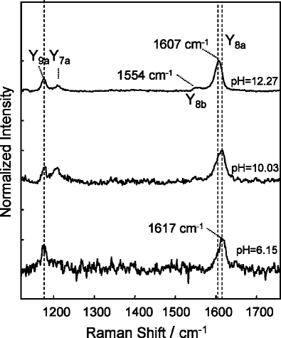
Resonance Raman spectra of TTR(105-115), in the wave-number range corresponding to vibrations of the phenolic side-chain of the tyrosines, excited at 239.5 nm, for three selected values of pH: vi.15, ten.03, and 12.27. Taken from Pieridou and Hayes (2009)
Plotting the frequency of the Y8a band equally a function of pH produces the titration curve. Plumbing fixtures the pH dependence to the Henderson–Hasselbach curve resulted in a pK a of x.2 ±0.2 for the peptide. This value represents an average value for the two Tyr in the peptide. The aforementioned method for free Tyr in solution gave ix.1 ±0.2.
Attempting to explain the higher pK a value for the TTR(105-115) peptide relative to complimentary Tyr, Pieridou and Hayes (2009) referred to microenvironments of the Tyr side-chains formed by the TTR(105-115) peptide. Tyr-105 occupies the N-terminal position and is expected to be fully exposed to solvent, hence its pK a should be similar to that of free Tyr. Tyr-114, on the other paw, is amidst residues with H-bonding side-bondage, Ser112 and Ser115, which can act either every bit proton donors or acceptors. In addition, the neighboring Pro-113 residuum may maintain some specific structure within the peptide fragment. Both of these factors may elevate the pK a of Tyr-114, and as a consequence the average pK a value determined in the experiment is as well elevated.
Conclusions and perspectives
We have discussed absorption of UV light by the side-concatenation of Tyr residues in proteins, equally well as several other spectroscopic backdrop of this chromophore, all valuable probes of protein construction.
Although large extinction coefficient, emission quantum yield, and high sensitivity to changes in the microenvironment have led to Trp being routinely used every bit an intrinsic marker in proteins, inclusion of Tyr in the arsenal of chromophores used for characterization of the protein construction has clear advantages. Phenol is more polar than indole, therefore Tyr should react more strongly to environmental changes than Trp. Shifts in the absorbance spectrum due to changes in the polarity of the environment are generally larger for Tyr than for tryptophan (Fornander et al. 2014).
The phenol motif in Tyr provides unique physicochemical properties and chemical reactivity that enables this amino acrid residue to achieve a plethora of biosynthetic transformations and molecular interactions (Jones et al. 2014).
The spectroscopic properties described above may be used to follow these transformations and gain insight into these interactions. Experiments described in this review may present useful hints for studies of structural properties of proteins, employing other possible experimental and/or theoretical methods because UV–Vis absorption and fluorescence spectrometers, as well equally their more specific developments of LD, CD, or resonance Raman scattering, are readily available, and information technology is easy to supplement an investigation with these spectroscopies.
References
-
Ahmad A, Millett IS, Doniach Southward, Uversky VN, Fink AL (2003). Biochemistry 42:11404
-
Aihara H, Ito Y, Kurumizaka H, Yokoyama South, Shibata T (1999). J Mol Biol 290:495
-
Aitken A, Learmonth MP (2009) Poly peptide protocols handbook. In: Walker JM (ed) Springer protocols handbooks. chap. 1. Humana Press, New York, pp 3–half-dozen
-
Baumann P, West SC (1998). Trends Biochem Sci 23:247
-
Bekard IB, Dunstan DE (2009). Biophys J 97:2521
-
Breydo LP, Shevchenko AA, Kost OA (1997). Russ Chem Bull 46:1339
-
Chatani Due east, Hayashi R, Moriyama H, Ueki T (2002). Protein Sci 11 :72
-
Conway AB, Lynch TW, Zhang Y, Fortin GS, Fung CW, Symington LS, Rice PA (2004). Nat Struct Mol Biol 11:791
-
Couling VW, Foster NW, Klenerman D (1997). J Raman Spectr 28:33
-
Cox MM (2009). Proc Natl Acad Sci United states 106:13147
-
Crammer JL, Neuberger A (1943). Biochem J 37:302
-
Edelhoch H, Perlman RL, Wilchek M (1969). Ann New York Acad Sci 158:391
-
Edelhoch H, Lippoldt RE (1969). J Biol Chem 244:3876
-
Fersht AR (1993). FEBS Lett 325:5
-
Fornander LH, Feng B, Beke-Somfai T, Nordén B (2014) J Phys Chem B 118:9247
-
Georgieva DN, Genov N, Rajashankar KR, Aleksiev B, Betzel C (1999). Spectrchim Acta Part A 55:239
-
Goldfarb AR, Saidel LJ, Mosovich E (1951) J Biol Chem 193:397
-
Gorbunoff MJ (1967). Biochemistry half-dozen:1606
-
Gustavsson A, Engstrom U, Westermark P (1991). Biochem Biophys Res Commun 175:1159
-
Hermans J Jr (1962). Biochemistry one:193
-
Horwitz J, Strickland EH, Billups C (1970). J Amer Chem Soc 92:2119
-
Howlin B, Moss DS, Harris GW (1989). Acta Crystallogr 45:851
-
Hua Q (2010). Poly peptide Prison cell 1:537
-
Jones LH, Narayanan A, Hett EC (2014). Molec BioSys 10:952
-
Kueltzo LA, Middaugh CR (2005) Methods for structural analysis of protein pharmaceuticals. In: Jiskoot W, Crommelin DJA (eds), vol iii. AAPS Printing, Arlington, pp 1–26
-
Kuwajima K, Ogawa Y, Sugai Southward (1979). Biochemistry xviii:878
-
Leydier C, Clottes E, Couthon F, Marcillat O, Vial C (1997). Biochem Mol Biol International 41:777
-
Lakowicz JR, Kierdaszuk B, Callis P, Malak H, Gryczynski I (1995). Biophys Chem 56:263
-
Larkin P (2011) Infrared and Raman spectroscopy; principles and spectral interpretation. Elsevier, Waltham, p 02451
-
Lucas LH, Ersoy BA, Kueltzo LA, Joshi SB, Brandau DT, Thyagarajapuram Due north, Peek LJ, Middaugh CR (2006). Prot Sci fifteen:2228
-
Matouschek A, Serrano Fifty, Fersht AR (1992). J Mol Biol 224:819
-
Mihashi One thousand, Ooi T (1965). Biochemistry four:805
-
Nordén B, Elvingson C, ad MK, Sjoberg B, Ryberg H, Ryberg G, Moztensen K, Takahashi 1000 (1992). J Mol Biol 226:1175
-
Nordlund TM, Liu XY, Sommer JH (1986) Proc. Natl Acad Sci USA 83:8977
-
Pellegrini 50, Yu DS, Lo T, Anand Southward, Lee M, Blundell TL, Venkitaraman AR (2002). Nature 420:287
-
Pieridou GK, Hayes SC (2009) Phys. Chem Chem Phys xi:5302
-
Platzer G, Okon M, McIntosh LP (2014). J Biomolec NMR 60:109
-
Ragone R, Colonna G, Balestrieri C, Servillo L, Irace G (1984). Biochemistry 23:1871
-
Reymer A, Frykholm K, Morimatsu K, Takahashi Yard, Nordén B (2009). Proc Natl Acad Sci Us 106:13248
-
Servillo L, Colonna G, Balestrieri C, Ragone R, Irace One thousand (1982). Anal Biochem 126:251
-
Shugar D (1952). Biochem. J 52:142
-
Steiner RF, Edelhoch H (1963). J Biol Chem 238:925
-
Simmons NS, Glazer AN (1967). J Amer Chem Soc 89:5040
-
Simpson RT, Vallee BL (1966). Biochemistry five:2531
-
Tanford C, Hauenstein JD, Rands DG (1956). J Amer Chem Soc 77:6409
-
VanderMeulen DL, Govindjee (1977). Eur J Biochem 78:585
-
Vieira M, Saraiv MJ (2014). Biomol Concepts 5:45
-
Wilson EB Jr. (1934). Phys Rev 45:706
-
Woody AM, Woody RW (2003). Biopolymers (Biospectroscopy) 72:500
Acknowledgments
Our piece of work was supported by the University of Warsaw (JMA, grant BST-166700), and by the Institute of Biochemistry & Biophysics, Smooth Academy of Sciences (DS).
Open Access
This article is distributed nether the terms of the Creative Commons Attribution iv.0 International License (http://creativecommons.org/licenses/past/4.0/), which permits unrestricted use, distribution, and reproduction in whatsoever medium, provided you give appropriate credit to the original author(s) and the source, provide a link to the Artistic Eatables license, and indicate if changes were made.
Author data
Affiliations
Corresponding author
Ethics declarations
Conflict of interests
Jan M. Antosiewicz declares that he has no conflict of interest. David Shugar declares that he has no conflict of interest.
Additional data
Ethical approval
This commodity does not contain any studies with human participants or animals performed by any of the authors.
Rights and permissions
Open up Access This article is distributed nether the terms of the Creative Commons Attribution 4.0 International License (http://creativecommons.org/licenses/by/4.0/), which permits unrestricted use, distribution, and reproduction in whatever medium, provided you lot requite advisable credit to the original author(southward) and the source, provide a link to the Creative Commons license, and betoken if changes were fabricated.
Reprints and Permissions
Near this article
Cite this article
Antosiewicz, J.Chiliad., Shugar, D. UV–Vis spectroscopy of tyrosine side-groups in studies of protein structure. Role ii: selected applications. Biophys Rev viii, 163–177 (2016). https://doi.org/10.1007/s12551-016-0197-7
-
Received:
-
Accustomed:
-
Published:
-
Consequence Date:
-
DOI : https://doi.org/ten.1007/s12551-016-0197-7
Keywords
- Tyrosine
- UV–Vis assimilation
- Fluorescence
- Linear and circular dichroism
- Resonance Raman handful
Source: https://link.springer.com/article/10.1007/s12551-016-0197-7
0 Response to "How to Read Uv Absorption Spectrum Ribosomes"
Postar um comentário